
Appendix (anatomy)
| Appendix | |
|---|---|
 Appendix with surrounding structures | |
 Variations of the appendix | |
| Details | |
| Precursor | Midgut |
| System | Digestive system |
| Artery | Appendicular artery |
| Vein | Appendicular vein |
| Identifiers | |
| MeSH | D001065 |
| TA98 | A05.7.02.007 |
| TA2 | 2976 |
| FMA | 14542 |
| Anatomical terminology | |
The appendix (pl.: appendices or appendixes; also vermiform appendix; cecal (or caecal, cæcal) appendix; vermix; or vermiform process) is a finger-like, blind-ended tube connected to the cecum, from which it develops in the embryo.
The cecum is a pouch-like structure of the large intestine, located at the junction of the small and the large intestines. The term "vermiform" comes from Latin and means "worm-shaped". In the early 2000s the appendix was reassessed and is no longer considered a vestigial organ.[1][2] The appendix may serve as a reservoir for beneficial gut bacteria.
Structure
[edit]The human appendix averages 9 mm (0.35 in) in length, ranging from 5 to 35 mm (0.20 to 1.38 in). The diameter of the appendix is 6 mm (0.24 in), any larger is considered a thickened or inflamed appendix. The appendix is usually located in the lower right quadrant of the abdomen, near the right hip bone. The base of the appendix is located 2 cm (0.79 in) beneath the ileocecal valve that separates the large intestine from the small intestine. Its position within the abdomen corresponds to a point on the surface known as McBurney's point.
The appendix is connected to the mesentery in the lower region of the ileum, by a short region of the mesocolon known as the mesoappendix.[3]
Variation
[edit]Some identical twins–known as mirror image twins–can have a mirror-imaged anatomy, a congenital condition with the appendix located in the lower left quadrant of the abdomen instead of the lower right.[4][5] Intestinal malrotation may also cause displacement of the appendix to the left side.
While the base of the appendix is typically located 2 cm (0.79 in) below the ileocecal valve, the tip of the appendix can be variably located–in the pelvis, outside the peritoneum or behind the cecum.[6] The prevalence of the different positions varies amongst populations for example, the retrocecal position is most common in Ghana and Sudan, with 67.3% and 58.3% occurrence respectively, in comparison to Iran and Bosnia where the pelvic position is most common, with 55.8% and 57.7% occurrence respectively.[7][8][9][10]
In rare cases, the appendix may not be present at all (laparotomies for suspected appendicitis have given a frequency of 1 in 100,000).[11]
Sometimes a semi-circular fold of mucous membrane appears at the opening of the appendix. This valve of the vermiform appendix is also called Gerlach's valve.[3]
Functions
[edit]Maintaining gut flora
[edit]
Gut-associated lymphoid tissue is immune tissue surrounding the appendix and appearing elsewhere in the gut. It carries out several important functions. Initially, the appendix's distinctive shape and its apparent lack of specific function was judged by an absence of side effects following its removal.[12]
William Parker, Randy Bollinger, and colleagues at Duke University proposed in 2007 that the appendix serves as a haven for useful bacteria when illness or antibiotics flushes the bacteria from the intestines.[13][14] This proposition is based on an understanding of how the immune system supports the growth of beneficial intestinal bacteria,[15][16] in combination with other well-known features of the appendix, including its architecture, its location just below the normal one-way flow of food and germs in the large intestine, and its association with copious amounts of immune tissue.
Research in 2012 reported that individuals without an appendix were four times as likely to have a recurrence of Clostridioides difficile colitis.[17] The appendix, therefore, may act as a reservoir for beneficial bacteria.[13] This reservoir could repopulate the gut flora following a bout of gastrointestinal illness.[14]
Immune and lymphatic systems
[edit]The appendix is an important component of mammalian mucosal immune function, particularly B cell-mediated immune responses and extrathymically derived T cells. This structure helps in the proper movement and removal of waste matter in the digestive system, contains lymphatic vessels that regulate pathogens, and lastly, might even produce early defence against disease. Additionally, this may provide immune defence from invading pathogens and stimulating B and T cells to fight viruses and bacteria that infect that portion of the bowel and training them so that immune responses are targeted and more able to fight pathogens.[18] In addition, immune cells called innate lymphoid cells help the appendix maintain digestive health.[19]
A 2016 study reported a positive correlation between the presence of the appendix and the concentration of cecal lymphoid tissue, which supports the suggestion that the appendix has major immune benefits.[20]
Disease
[edit]
Common diseases of the appendix (in humans) are appendicitis and carcinoid tumors (appendiceal carcinoid).[21] Appendix cancer accounts for about 1 in 200 of all gastrointestinal malignancies. In rare cases, adenomas are present.[22]
Appendicitis
[edit]Appendicitis is a condition characterized by inflammation of the appendix. Pain often begins in the center of the abdomen, corresponding to the appendix's development as part of the embryonic midgut. This pain is typically a dull, poorly localized, visceral pain.[23]
As the inflammation progresses, the pain begins to localize more clearly to the right lower quadrant, as the peritoneum becomes inflamed. This inflammation, or peritonitis, results in rebound tenderness (pain upon removal of pressure rather than the application of pressure). In particular, it presents at McBurney's point, 1/3 of the way along a line drawn from the anterior superior iliac spine to the umbilicus. Typically, point (skin) pain is not present until the parietal peritoneum is inflamed as well. Fever and an immune system response are also characteristic.[23] Other signs and symptoms include nausea and vomiting, fever, constipation or diarrhea, abdominal bloating, or flatulence.[24]
Untreated, the appendix may rupture, leading to peritonitis, followed by shock, and, if still untreated, death.[23]
Appendectomy is the surgical removal of the appendix. This removal is typically performed as an emergency procedure in a suffering from acute appendicitis. In the absence of surgical facilities, intravenous antibiotics are used to delay or avoid the onset of sepsis. In some cases, the appendicitis resolves; otherwise, an inflammatory mass forms around the appendix. This is a relative contraindication to surgery.
Uses
[edit]The appendix can be used to construct an efferent urinary conduit, in an operation known as the Mitrofanoff procedure,[25] in people with a neurogenic bladder.
The appendix can also be used to access the colon in children with paralysed bowels or major sphincter dysfunction. The appendix is lifted from the abdomen and a catheter can be attached which allows the colon to be irrigated (via normal defaecation).[26]
History
[edit]Charles Darwin suggested that the appendix was mainly used by earlier hominids for digesting fibrous vegetation, before evolving. The large cecum of some herbivorous animals, such as the horse, rodents or the koala, appears to support this hypothesis. The koala's cecum enables it to host bacteria that specifically help to break down cellulose. Human ancestors may have also relied upon to digest a diet rich in foliage.
As people shifted to eat more easily digested foods, the cecum became less necessary for digestion. Mutations that were previously deleterious lost their salience, which allowed them to survive. These alleles became more frequent and the cecum shrank. After millions of years, the human cecum became the appendix.[27]
Dr. Heather F. Smith and colleagues stated:
Recently ... improved understanding of gut immunity has merged with current thinking in biological and medical science, pointing to an apparent function of the mammalian cecal appendix as a safe-house for symbiotic gut microbes, preserving the flora during times of gastrointestinal infection in societies without modern medicine. This function is potentially a selective force for the evolution and maintenance of the appendix. Three morphotypes of cecal-appendices can be described among mammals based primarily on the shape of the cecum: a distinct appendix branching from a rounded or sac-like cecum (as in many primate species), an appendix located at the apex of a long and voluminous cecum (as in the rabbit, greater glider and Cape dune mole rat), and an appendix in the absence of a pronounced cecum (as in the wombat). In addition, long narrow appendix-like structures are found in mammals that either lack an apparent cecum (as in monotremes) or lack a distinct junction between the cecum and appendix-like structure (as in the koala). A cecal appendix has evolved independently at least twice and represents yet another example of convergence in morphology between Australian marsupials and placentals in the rest of the world. Although the appendix has been lost by numerous species, it has also been maintained for more than 80 million years in at least one clade.[28]
A 2013 study estimated that the appendix independently evolved in different animals 32-38 times but to have been lost no more than six times.[29] A 2017 study of an updated database yielded similar results, with estimates of at least 29 gains and at most 12 losses (all of which were ambiguous).[30] This suggests that the cecal appendix has a selection advantage and is not vestigial. It appears to be associated with greater longevity.[31] For example, a 2023 study reported protective functions against diarrhea in young primates.[32][33]
That function may be particularly useful in the absence of sanitation and healthcare, where diarrhea may be prevalent. Epidemiological data on the cause of death in developing countries collected by the World Health Organization in 2001 reported that acute diarrhea was the fourth leading cause of disease-related death in developing countries. Two of the other leading causes of death were claimed to have exerted limited or no selection pressure.[34]
Gallery
[edit]-
 Abdominal ultrasound showing a normal appendix between the external iliac artery and the abdominal wall
 -
 Illustration depicting the location of the appendix in a child
 -
 Normal location of the appendix relative to other organs of the digestive system (frontal view)
 -
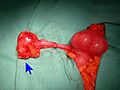
 Vermiform appendix
 -
 Mucinous adenocarcinoma of the appendix tip
 -
-
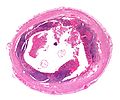
 Micrograph of entry point of appendicular arteries (arrows at level of inner muscular layer), not to be confused with a perforation.


See also
[edit]- Meckel's diverticulum
- Appendix of the epididymis, a detached efferent duct of the epididymis
- Appendix testis, a vestigial remnant of the Müllerian duct
- Epiploic appendix, one of several small pouches of fat on the peritoneum along the colon and rectum
- Appendix of the laryngeal ventricle, a sac that extends from the laryngeal ventricle
- Mesoappendix, the portion of the mesentery that connects the ileum to the vermiform appendix
References
[edit]- ^ Kooij IA, Sahami S, Meijer SL, Buskens CJ, Te Velde AA (October 2016). "The immunology of the vermiform appendix: a review of the literature". Clinical and Experimental Immunology. 186 (1): 1–9. doi:10.1111/cei.12821. PMC 5011360. PMID 27271818.
- ^ Smith, H. F.; Fisher, R. E.; Everett, M. L.; Thomas, A. D.; Randal Bollinger, R.; Parker, W. (2009). "Comparative anatomy and phylogenetic distribution of the mammalian cecal appendix". Journal of Evolutionary Biology. 22 (10): 1984–1999. doi:10.1111/j.1420-9101.2009.01809.x. PMID 19678866.
- ^ a b Golalipour, M.J.; Arya, B.; Jahanshahi, M.; Azarhoosh, R. (2003). "Anatomical Variations Of Vermiform Appendix In South-East Caspian Sea (Gorgan-IRAN)" (PDF). J. Anat. Soc. India. Archived (PDF) from the original on 11 July 2020. Retrieved 1 October 2014.
- ^ "Unusual Types of Twins". Multiples of America. Archived from the original on 2 May 2014. Retrieved 30 April 2014.
- ^ Gedda L, Sciacca A, Brenci G, Villatico S, Bonanni G, Gueli N, Talone C (1984). "Situs viscerum specularis in monozygotic twins". Acta Geneticae Medicae et Gemellologiae. 33 (1): 81–5. doi:10.1017/S0001566000007546. PMID 6540028.
- ^ Paterson-Brown, S. (2007). "15. The acute abdomen and intestinal obstruction". In Parks, Rowan W.; Garden, O. James; Carter, David John; Bradbury, Andrew W.; Forsythe, John L. R. (eds.). Principles and practice of surgery (5th ed.). Edinburgh: Churchill Livingstone. ISBN 978-0-443-10157-1.
- ^ Clegg-Lamptey JN, Armah H, Naaeder SB, Adu-Aryee NA (December 2006). "Position and susceptibility to inflammation of vermiform appendix in Accra, Ghana". East African Medical Journal. 83 (12): 670–3. doi:10.4314/eamj.v83i12.9498. PMID 17685212.
- ^ Bakheit MA, Warille AA (June 1999). "Anomalies of the vermiform appendix and prevalence of acute appendicitis in Khartoum". East African Medical Journal. 76 (6): 338–40. PMID 10750522.
- ^ Ghorbani A, Forouzesh M, Kazemifar AM (2014). "Variation in Anatomical Position of Vermiform Appendix among Iranian Population: An Old Issue Which Has Not Lost Its Importance". Anatomy Research International. 2014: 313575. doi:10.1155/2014/313575. PMC 4176911. PMID 25295193.
- ^ Denjalić A, Delić J, Delić-Custendil S, Muminagić S (2009). "[Variations in position and place of formation of appendix vermiformis found in the course of open appendectomy]". Medicinski Arhiv (in Bosnian). 63 (2): 100–1. PMID 19537667.
- ^ Zetina-Mejía CA, Alvarez-Cosío JE, Quillo-Olvera J (2009). "Congenital absence of the cecal appendix. Case report". Cirugia y Cirujanos. 77 (5): 407–10. PMID 19944032.
- ^ Kumar, Vinay; Robbins, Stanley L.; Cotran, Ramzi S. (1989). Robbins' pathologic basis of disease (4th ed.). Philadelphia: Saunders. pp. 902–3. ISBN 978-0-7216-2302-3.
- ^ a b "Scientists may have found appendix's purpose". NBC News. Associated Press. 5 October 2007. Archived from the original on 4 February 2020. Retrieved 24 August 2019.
- ^ a b Randal Bollinger R, Barbas AS, Bush EL, Lin SS, Parker W (December 2007). "Biofilms in the large bowel suggest an apparent function of the human vermiform appendix". Journal of Theoretical Biology. 249 (4): 826–31. Bibcode:2007JThBi.249..826R. doi:10.1016/j.jtbi.2007.08.032. PMID 17936308.
- ^ Sonnenburg JL, Angenent LT, Gordon JI (June 2004). "Getting a grip on things: how do communities of bacterial symbionts become established in our intestine?". Nature Immunology. 5 (6): 569–73. doi:10.1038/ni1079. PMID 15164016. S2CID 25672527.
- ^ Everett M.L.; Palestrant D.; Miller S.E.; Bollinger R.R.; Parker W. (2004). "Immune exclusion and immune inclusion: a new model of host-bacterial interactions in the gut". Clinical and Applied Immunology Reviews. 4 (5): 321–32. doi:10.1016/j.cair.2004.03.001.
- ^ Dunn, Rob (January 2, 2012). "Your Appendix Could Save Your Life". Scientific American. Archived from the original on 11 November 2020. Retrieved 22 December 2016.
- ^ Zahid A (April 2004). "The vermiform appendix: not a useless organ". Journal of the College of Physicians and Surgeons--Pakistan. 14 (4): 256–8. PMID 15228837.
- ^ Rankin LC, Girard-Madoux MJ, Seillet C, Mielke LA, Kerdiles Y, Fenis A, et al. (February 2016). "Complementarity and redundancy of IL-22-producing innate lymphoid cells". Nature Immunology. 17 (2): 179–86. doi:10.1038/ni.3332. PMC 4720992. PMID 26595889.
- ^ Smith H, Parker W, Kotze S, Laurin M (September 2016). "Morphological evolution of the mammalian cecum and cecal appendix". Comptes Rendus Palevol. 16 (1): 39–57. doi:10.1016/j.crpv.2016.06.001.
- ^ "Miscellaneous conditions of the appendix". Seminars in Diagnostic Pathology. 21 (2). National Institutes of Health: 151–63. 2004. doi:10.1053/j.semdp.2004.11.006. PMID 15807474.
- ^ "Statistics about Appendix disorder". rightdiagnosis.com. Archived from the original on 2019-10-16. Retrieved 2020-12-30.
- ^ a b c Miller R., Kenneth; Levine, Joseph (2002). Biology. Prentice Hall. pp. 92–98. ISBN 978-0-13-050730-3.
- ^ "Appendicitis - Symptoms and causes - Mayo Clinic". mayoclinic.org. Mayo Clinic. Archived from the original on 25 November 2020. Retrieved 29 December 2020.
- ^ Mingin GC, Baskin LS (2003). "Surgical management of the neurogenic bladder and bowel". International Braz J Urol. 29 (1): 53–61. doi:10.1590/S1677-55382003000100012. PMID 15745470.
- ^ "Wellington Children's Hospital: Caring for an ACE or Chait Tube: Healthpoint". Archived from the original on 16 October 2019. Retrieved 22 December 2016.
- ^ Darwin, Charles (1871). "Jim's Jesus". The Descent of Man, and Selection in Relation to Sex. London: John Murray.
- ^ Smith HF, Fisher RE, Everett ML, Thomas AD, Bollinger RR, Parker W (October 2009). "Comparative anatomy and phylogenetic distribution of the mammalian cecal appendix". Journal of Evolutionary Biology. 22 (10): 1984–99. doi:10.1111/j.1420-9101.2009.01809.x. PMID 19678866. S2CID 6112969.
- ^ Smith H. F.; Parker W.; Kotzé, S. H.; Laurin, M. (2013). "Multiple independent appearances of the cecal appendix in mammalian evolution and an investigation of related ecological and anatomical factors". Comptes Rendus Palevol. 12 (6): 339–354. Bibcode:2013CRPal..12..339S. doi:10.1016/j.crpv.2012.12.001.
- ^ Smith H. F.; Parker W.; Kotzé, S. H.; Laurin, M. (2017). "Morphological evolution of the mammalian cecum and cecal appendix". Comptes Rendus Palevol. 11 (1): 39–57. Bibcode:2017CRPal..16...39S. doi:10.1016/j.crpv.2016.06.001.
- ^ Collard, Maxime K.; Bardin, Jérémie; Laurin, Michel; Ogier-Denis, Eric (November 2021). "The cecal appendix is correlated with greater maximal longevity in mammals". Journal of Anatomy. 239 (5): 1157–1169. doi:10.1111/joa.13501. ISSN 0021-8782. PMC 8546507.
- ^ Collard, Maxime K.; Bardin, Jérémie; Marquet, Bertille; Laurin, Michel; Ogier-Denis, Éric (2023-09-23). "Correlation between the presence of a cecal appendix and reduced diarrhea severity in primates: new insights into the presumed function of the appendix". Scientific Reports. 13 (1): 15897. Bibcode:2023NatSR..1315897C. doi:10.1038/s41598-023-43070-5. ISSN 2045-2322. PMC 10517977. PMID 37741857.
- ^ Laurin M, Everett ML, Parker W (April 2011). "The cecal appendix: one more immune component with a function disturbed by post-industrial culture". Anatomical Record. 294 (4): 567–79. doi:10.1002/ar.21357. PMID 21370495. S2CID 3237168.
- ^ "Evolution of the Appendix: A Biological 'Remnant' No More". Duke Medicine News and Communications. 21 August 2009 [20 August 2009]. Archived from the original on 2012-07-26.
Further reading
[edit]- Appendix May Actually Have a Purpose–2007 WebMD article
- Anatomy photo:37:12-0102 at the SUNY Downstate Medical Center–"Abdominal Cavity: The Cecum and the Vermiform Appendix"
- "The vestigiality of the human vermiform appendix: A Modern Reappraisal"–evolutionary biology argument that the appendix is vestigial
- Smith HF, Fisher RE, Everett ML, Thomas AD, Bollinger RR, Parker W (October 2009). "Comparative anatomy and phylogenetic distribution of the mammalian cecal appendix". Journal of Evolutionary Biology. 22 (10): 1984–99. doi:10.1111/j.1420-9101.2009.01809.x. PMID 19678866. S2CID 6112969.
- Cho, Jinny (August 27, 2009). "Scientists refute Darwin's theory on appendix". The Chronicle (Duke University). (News article on the above journal article.)
External links
[edit] Media related to Appendix (anatomy) at Wikimedia Commons